

Acute Myeloid Leukemia With Myelodysplasia-Related Features

Overview
Acute myeloid leukemia (AML) with myelodysplasia-related features includes those forms of AML that occur in patients with a history of a myelodysplastic syndrome (MDS) or a myelodysplastic/myeloproliferative neoplasm (MDS/MPN); it also includes those forms of AML with morphologic features or cytogenetic abnormalities characteristic of an MDS.
Disorders
- AML arising in MDS or in MDS/MPN
- AML with multilineage dysplasia or with MDS-related cytogenetic abnormalities
Definition
By definition, the diagnosis of AML with myelodysplasia-related features requires the presence of at least 20% blasts in the peripheral blood (PB) or bone marrow (BM), an absence of any of the recurrent cytogenetic abnormalities seen in AML, and the absence of a history of cytotoxicity or radiation therapy for an unrelated disease.
Epidemiology
This category of AML primarily occurs in elderly patients; it is rare in children. AML with multilineage dysplasia represents 25-35% of AML cases.1,2,3,4
Clinical Features
Patients with this category of AML often present with severe pancytopenia. Cases arising in association with an MDS or MDS/MPN may be slowly progressive.
Morphologic Features
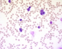
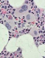
AML with multilineage dysplasia is established by demonstrating at least 50% of cells in at least two BM cell lines (ie, dysgranulopoiesis, dyserythropoiesis, or dysmegakaryopoiesis). Features of dysgranulopoiesis include hypogranulosis, hyposegmented neutrophils (ie, pseudo-Pelger Huet forms), or abnormally segmented nuclei (seeMedia files 1-3). As such, cytochemical staining, especially with myeloperoxidase (MPO), may be aberrant, because patients may develop an acquired MPO deficiency as part of the dysplastic process.5,6,7
Dysgranulopoiesis: Hypogranulosis.
Dysgranulopoiesis: Vacuolization. Image courtesy of Rector and Visitors of the University of Virginia.
Dysgranulopoiesis demonstrating Chediak-Higashi-like granules (arrow).
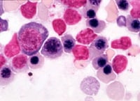
Features of dyserythropoiesis include megaloblastoid forms; nuclear budding; nuclear irregularities; nuclear fragmentation and multinucleation; karyorrhexis; mitoses within erythroid forms; ringed sideroblasts; and cytoplasmic vacuolization (see Media file 4).
Dyserythropoiesis.
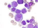
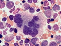
Features of dysmegakaryopoiesis include micro-megakaryocytic forms, as well as mononucleated and multinucleated forms (see Media files 5-6).
Dysmegakaryopoiesis: Mononucleated form.
Dysmegakaryopoiesis: Multinucleated form.
Immunophenotypic Features and Methods
The maturing myeloid cells in MDS may show immunophenotypic abnormalities by flow cytometric immunophenotyping, as follows: a decrease in side light scatter properties (due to hypogranulosis), increased intensity of CD33 expression, persistence of CD33 to later myeloid stages, decreased CD10 expression, abnormal patterns of CD11b versus CD16 expression or CD13 versus CD16 expression, deviations in myeloid antigen intensity, dyssynchronous expression of 2 myeloid antigens, and aberrant nonmyeloid antigen expression. The blasts in MDS may show anomalous expression of CD56 and CD7.8 There is usually an increase in multidrug resistance glycoprotein (MDR-1) on the cell surface.9,10,11 By immunohistochemical staining with CD34 and/or CD117, MDS often shows a cluster of blasts interstitially.
Molecular/Genetic Features and Methods
The cytogenetic abnormalities that are considered to be MDS related and that are sufficient to categorize such AML as MDS related in the absence of a history of an MDS or MDS/MPN and even in the absence of demonstrable multilineage dysplasia by morphologic criteria include the following:
- Complex karyotype (at least 3 unrelated abnormalities, none of which include the recurrent cytogenetic abnormalities encountered in AML)
- Unbalanced abnormalities [-7/del(7q), -5/del(5q), i(17q)/t(17p), -13/del(13q), del(11q), del(12p)/t(12p), del(9q), and idic(X)(q13)]
- Balanced abnormalities [t(11;16)(q23;p13.3), t(3;21)(q26.2;q21.2), t(1;3)(p36.3;q21.1), t(2;11)(p21;q23), t(5;12)(q33;p12), t(5;7)(q33;q11.2), t(5;17)(q33;p13), t(5;10)(q33;q21), and t(3;5)(q25;q34)]
Prognosis and Predictive Factors
Patients with this category of AML generally have a poor prognosis; the percentage of patients who achieve complete remission is lower with this type of AML than with other AML types. As mentioned above, cases arising in association with an MDS or MDS/MPN may show slower progression and thus may be less clinically aggressive. The prognosis is dependent on the associated cytogenetic abnormalities; high-risk cytogenetic abnormalities include the following: -5, -7, del(5q), abn(3q), and a complex karyotype.
DIFFERENTIALS
Multimedia
 | Media file 1: Dysgranulopoiesis: Hypogranulosis. |
 | Media file 2: Dysgranulopoiesis: Vacuolization. Image courtesy of Rector and Visitors of the University of Virginia. |
 | Media file 3: Dysgranulopoiesis demonstrating Chediak-Higashi-like granules (arrow). |
 | Media file 4: Dyserythropoiesis. |
 | Media file 5: Dysmegakaryopoiesis: Mononucleated form. |
 | Media file 6: Dysmegakaryopoiesis: Multinucleated form. |
Keywords
acute myeloid leukemia with myelodysplasia-related features, AML with myelodysplasia-related features, acute myeloid leukemia, AML, myelodysplasia, cytogenetic abnormalities

0 comments:
Post a Comment