

Anomalous Left Coronary Artery From the Pulmonary Artery: Multimedia

Multimedia
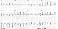 | Media file 1: Preoperative electrocardiogram in a 2-month-old infant with anomalous origin of the left coronary artery from the pulmonary artery demonstrating pathologic Q waves in leads I and aVL and diffuse ST-T wave changes consistent with an anterolateral infarction. |
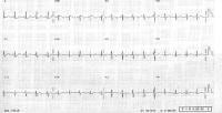 | Media file 2: Electrocardiogram in 2-month-old infant with anomalous origin of the left coronary artery from the pulmonary artery 17 months following successful surgical revascularization, demonstrating complete resolution of the anterolateral infarction pattern and ST-T wave changes. |
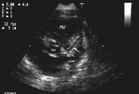 | Media file 3: Two-dimensional echocardiographic image (parasternal short axis view) in a patient with anomalous origin of the left coronary artery arising from the pulmonary artery (ALCAPA). The left coronary artery (white arrow) appears to course towards the main pulmonary artery (MPA) just above the pulmonary valve and not to the aortic root (Ao). RV = Right ventricle. |
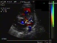 | Media file 4: Two-dimensional echocardiographic image with color flow mapping (parasternal short axis view) in the same patient with anomalous origin of the left coronary artery arising from the pulmonary artery (ALCAPA). The addition of color flow mapping to the 2-dimensional image demonstrates abnormal flow reversal within the left coronary artery (white arrows) towards the main pulmonary artery (MPA) just above the pulmonary valve. RV = Right ventricle. Ao = Aortic root. |
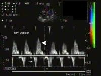 | Media file 5: Doppler interrogation of the abnormal color flow jet is depicted, demonstrating abnormal flow within the main pulmonary artery towards the transducer in diastole, which represents runoff from the anomalous left coronary artery (large white arrowhead). Small white arrow: Normal antegrade main pulmonary artery flow in systole. MPA = Main pulmonary artery. |
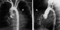 | Media file 6: Aortogram in a patient with suspected anomalous origin of the left coronary artery from the pulmonary artery (ALCAPA). Frontal (left panel) and lateral (right panel) images demonstrating an enlarged right coronary artery (small white arrow), which fills a small left coronary system (solid arrow head) via collaterals with eventual faint opacification of the main pulmonary artery (not demonstrated in this frame). |
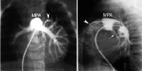 | Media file 7: Main pulmonary artery angiogram demonstrating the technique of stop flow angiography. There is retrograde opacification of the entire left coronary artery system, which originates from the distal main pulmonary artery (MPA), including the anterior descending (solid white arrowhead) and circumflex (small white arrow) branches. Left panel: Frontal image. Right panel: Lateral image. |

0 comments:
Post a Comment